Hyatt, J.G., Paterson, N.G., Devos, J.M., Oliviera, C.L.P., Jessen, C.M., Prevost, S., Hofmann, A., Pedersen, J.S., Winter, A. (2026)
Abstract
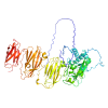
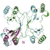
AAA proteases are hexameric ATP-dependent metallopeptidases that perform crucial proteolytic activities within prokaryotic and eukaryotic membranes. Structurally, protomers are comprised of catalytically active C-terminal domains that are anchored to the membrane by an N-terminal autonomous folding unit. In this study, we determined the fold, stability, and oligomeric state of the N-terminal intermembrane domains of human spastic paraplegia type 7 (SPG7)/ paraplegin protein and its bacterial orthologue FtsH using circular dichroism (CD), small-angle X-ray scattering (SAXS), small-angle neutron scattering (SANS) and X-ray crystallography. Solution-state analysis revealed that the N-terminal domain of paraplegin is a monomer in solution whereas FtsH forms a dimer. Unexpectedly, the N-terminal domain of paraplegin presents as a domain-swapped homodimer in our crystal structure that involves the first helix and first two beta-strands from one monomer and beta-strand 3, helix 2 and beta-strand 4 from another symmetry-related molecule. However, together they form an assembly which is similar to protomers observed for the N-terminal regions of FtsH and AfG3L2. Drawing from our structural data, we postulate that domain-swapping interactions of the N-terminal regions contribute to stability of the AAA protease hexamer containing paraplegin, demonstrating the extensive flexibility of the N-terminal portion of this protein and its role in achieving the appropriate molecular architecture required for function.